

It has been two centuries since Petrus Camper identified the superficial fascia and over 175 years since Sir Astley Cooper wrote his book "On the Anatomy of the Breast". In the 1990s, Ted Lockwood provided information on the importance of superficial layers of fascia in body contouring procedures. However, these descriptions cannot explain the three-dimensional fascial system of the breast.
In the article estet-portal.com you will be able to get acquainted in detail with the structure of the superficial fascia of the mammary gland, which is responsible for the shape of the breast.
Layer examination of the ligamentous apparatus of the mammary gland
Clinical observations have shown that there are two layers of superficial fascia that surround the chest. The body of the mammary gland is surrounded by an anterior and posterior layer of fatty tissue (Fig. 1).

Fig. 1. Anatomy of the ligamentous apparatus of the mammary gland
Starting from the surface of the chest and cutting deeper, we encounter 2 – 3 mm layer of subcutaneous fat under the dermis (Fig. 2). Next comes the superficial sheet of the superficial fascia (analogous to the Kramper fascia in the groin). This is followed by an anterior layer of adipose tissue 10 mm thick (Fig. 3), which covers the anterior surface of the mammary gland. The anterior adipose tissue layer is two to three times thicker than the posterior adipose tissue layer.
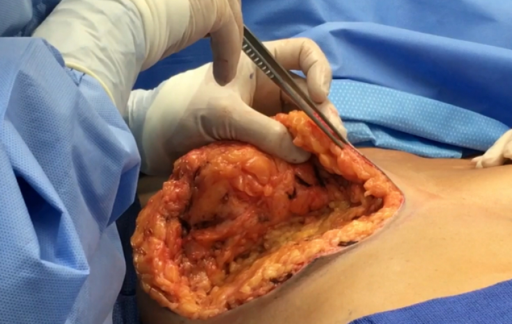
Fig. 2. Subcutaneous fat
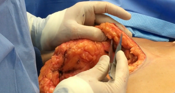
Fig. 3. Anterior fat layer
The superficial fascia consists of two sheets.
The superficial layer (Camper fascia) is thin and continues into the superficial fascia of the thigh. The deep leaflet (Scarpa's fascia) is well defined in the lower half of the abdomen.
The deeper sheet of fascia is several times thicker than the almost non-existent superficial sheet of superficial fascia.
Follow us on Facebook
Breast Ligament Apparatus: Mythbusting
In fact, the breast capsule (Fig. 4) is a pseudocapsule because there is no epithelium on its surface and it was formed as a result of compression by the surrounding fascia as the mammary gland expands during puberty.
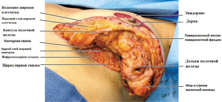
Fig. 4. Anatomy of the mammary gland
As a result of the fusion of the superficial and deep layers of the superficial fascia surrounding the chest, a "contour ligament" is formed; (Fig.4, Fig.5).
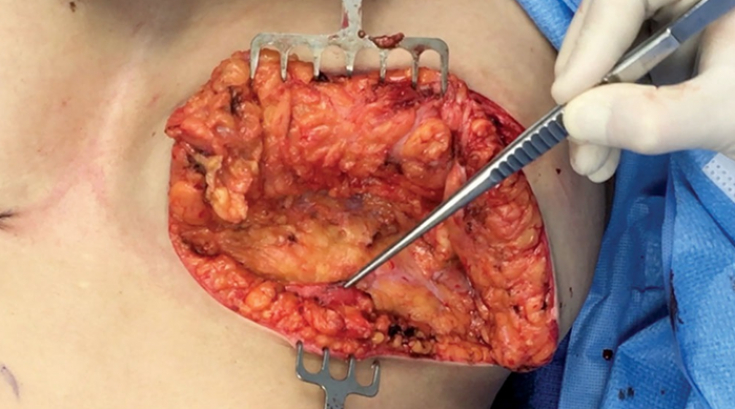
Fig. 5. Contour ligament formed as a result of the fusion of sheets of the superficial fascia
Modified vertical skin ligaments, Cooper's ligaments, originate from the deep sheet of the superficial fascia upward through the mammary gland (Fig. 6), penetrate into the anterior adipose tissue and superficial sheet of fascia, and terminate in the dermis.
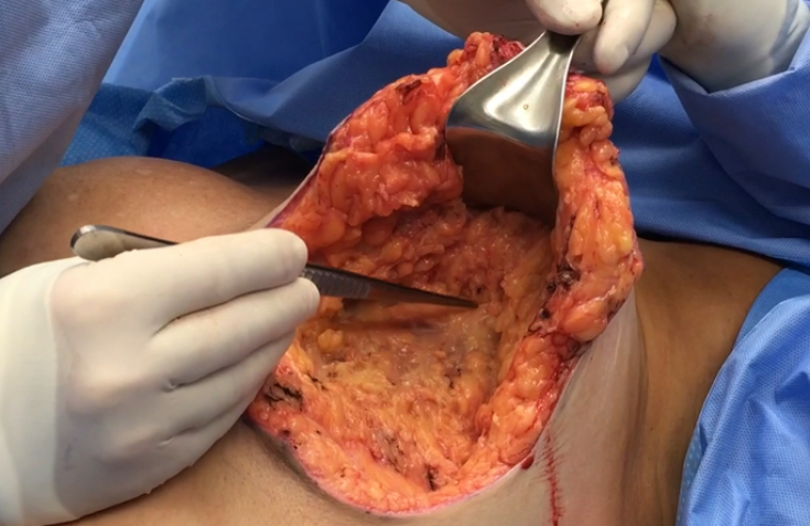
Fig. 6. Formation of Cooper's ligaments from a deep sheet of superficial fascia
The excretory ducts of the mammary gland enter through a round opening in the anterior fat layer and the superficial layer of the fascia under the areola (Fig. 8), which is called the annulus fibrosus (Fig. 9).
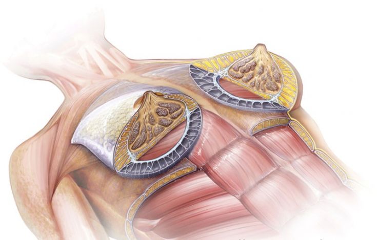
Fig. 8. Anatomy of the annulus
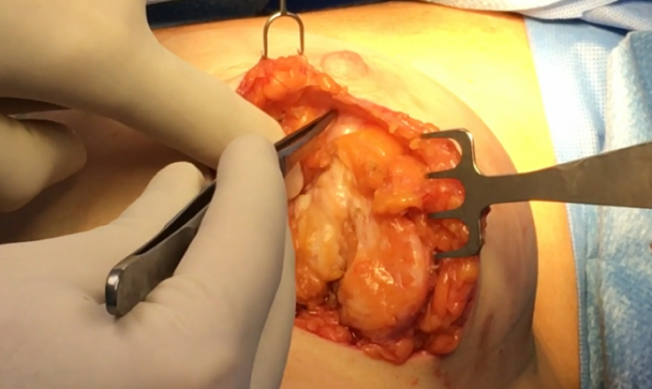
Fig. 9. Fibrous annulus
There is no anterior layer of adipose tissue and superficial fascia between the dermis of the areola and the mammary gland.
Vasily Khrapach: There is nothing better for a surgeon than active learning
The aesthetic value of the circular ligament
The circumferential ligament (Fig. 10) defines the perimeter of the breast. The circumferential ligament is a three-dimensional, roughly circular structure composed of superficial collagen fibers of the fascia that defines a ring of adipose tissue and attaches it to the deep fascia as a circular adhesion zone.
Circular ligament occurs in every patient, but differs in characteristics depending on the constitution.

Fig. 10. Location of the circular ligament
People with voluminous fat deposits have larger lobules of fatty tissue between the collagen fibers and therefore a larger ring of attachment.
The circumferential ligament fuses with the deep fascia that covers the chest wall and secures the breast in place; the subglandular space allows the movement of the breast and prevents its rigid attachment.
The structure of fat and fascia surrounding the mammary gland is responsible for the shape of the breast and is thus the key to breast aesthetics.
Anatomical features of the circular ligament
• The ball of connective tissue that forms the circular ligament varies in thickness, depending on the area.
• It has a high density of collagen fibers attached to the deep fascia covering the parasternal region and is therefore the area with the strongest attachment.
• In the lateral section, the collagen and fat of the circular ligament is less dense and covers a large area. For example, the circular ligament is very thin and contains virtually no fat in the area between the clavicular head of the pectoralis major muscle and its sternocostal head.
• The fascial cleft under the breast and within the borders of the circular ligament is known as the "subglandular" space and filled with loose connective tissue.
• Circular ligament provides access for nerves, lymphatics and large vessels. The main arterial supply comes from the chest wall and passes through the collagen fibers of the circular ligament before entering the chest (Figure 11).
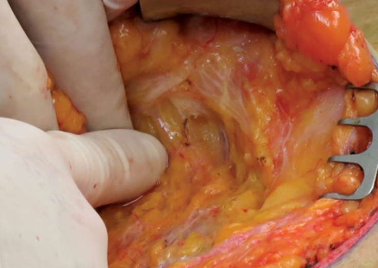
Fig. 11. Access of vessels and nerves through the circular ligament
The literature is full of conflicting results regarding the ligamentous apparatus of the breast, due to different access methods causing artifacts. This representation of the anatomy of the breast offers an opportunity to further explore the ontology of the breast and the dynamic forces that change the shape of the breast throughout life. Stretching and relaxation of the superficial fascial system due to aging or surgery leads to ptosis of the breast.
A thorough knowledge of breast anatomy is fundamental to both reconstructive and cosmetic breast surgery.
Thank you for staying with estet-portal.com. Read other interesting articles in the "Plastic Surgery" section. You might be interested in Anatomy of beauty: why patients choose anatomical implants for breast augmentation.
Based on Plastic and Reconstructive Surgery
Share:


Add a comment