

Breast diseases are one of the leading diseases in the world. Every year the number of patients with breast cancer increases, and postpartum inflammation of the breast becomes common.
Knowledge of the anatomical structure of the mammary glands and relationships with other organs is extremely important for the practitioner.
On estet-portal.com read about the structural features of the mammary glands, their blood supply, innervation and lymphatic system, as well as about the symptoms of possible breast pathologies.
The structure of the mammary gland: topography and main components of the organ
The breast is a modified sweat gland, which consists of glandular, adipose and connective tissue.
The structure of the mammary gland includes the following parts: the body of the breast (corpus mammae), the breast nipple (papilla mammaria) and the areola (areola).
At the periphery of the areola are modified sebaceous glands – tubercles of Montgomery.
The base of the mammary gland (glandula mammaria) is located at the level of 3-4 ribs on the surface of the pectoral fascia, which covers the pectoralis major muscle. The mammary gland is surrounded by a fatty capsule (capsula adiposa mammae).
The connective tissue cords that run from the thoracic fascia to the skin of the mammary gland are called the suspensory ligaments of the mammary gland.
Glandula mammaria consists of 16-20 cone-like lobes, the tops of which face the nipple.
The lobes of the gland by structure belong to complex alveolar-tubular glands, which are separated by loose connective and adipose tissues.
The lobes are built from 4-12 lobules of the mammary gland, consisting of alveoli (diameter 0.005-0.06 mm), from which the milk passages depart.
Follow us on Facebook
The structure of the mammary gland is based on the function of accumulation and excretion of nutrients during the lactation period.
Milk ducts are formed by the fusion of lobular and interlobular passages.
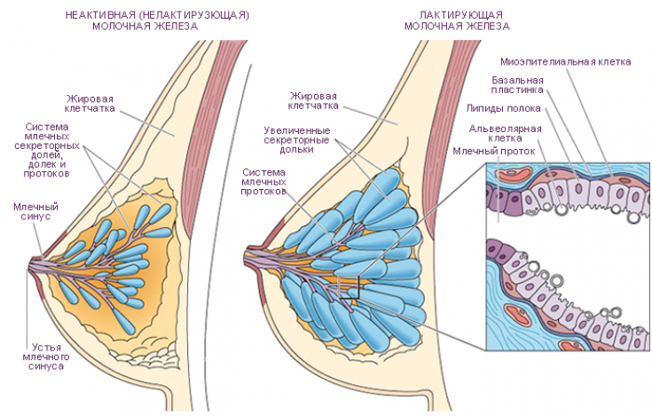
The milk ducts expand near the opening on the nipple and form the milk sinuses (sinus lactiferi).
The lactating mammary gland is characterized by changes in the size, consistency and ratio of tissues: the proportion of adipose tissue decreases, vascularization increases, and hypertrophy of the alveoli and ducts occurs. Nipple and areola receptors, spinal tracts, hypothalamus and pituitary gland form a reflex arc of the milk excretion mechanism. pledge of prolonged breastfeeding
Blood supply, innervation and lymphatic system of the breast
The blood supply to the mammary gland occurs due to the branches of the internal and lateral thoracic arteries, as well as the intercostal arteries.
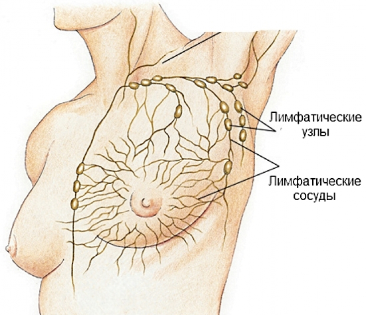 The main outflow of lymph from the mammary gland occurs towards the armpit and chest cavity.
The following lymph outflow tracts are distinguished from the breast:
• subclavian;
• parasternal;
• subclavian;• intercostal;
• retrosternal;
• cross
• Gerota's path (epigastric region).
The breast receives sensitive innervation from the lateral and medial branches of the mammary gland 2-4 and 4-6 intercostal nerves, respectively.
The sympathetic nerve fibers reach the breast along with blood vessels and sensory nerves.Female joys: breast asymmetry and the possibilities of its correction
Worrying symptoms: the clinical picture of breast pathologies
Most breast diseases can be diagnosed even in the initial stage of the pathology due to a vivid clinical picture and visible changes in the breast.
Breast diseases can be divided into 3 large groups:
1. inflammatory diseases (mastitis);
2. benign changes (mastopathy);
3. oncological diseases (intraductal, lobular cancer) shape of the gland, swollen lymph nodes, discharge from the nipples, changes in the color or structure of the skin of the breast, retraction of the nipple.Any unusual sensations and changes in the structure of the breast require careful clinical, instrumental and laboratory research.Thank you for staying with estet-portal.com. Read other interesting articles in the "Gynecology" section. You may also be interested in:
Share:


Add a comment